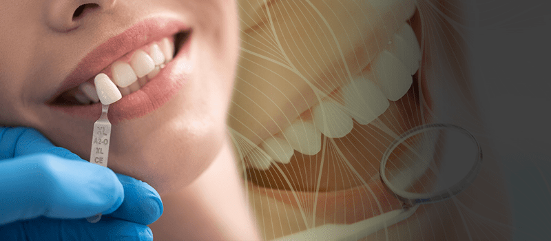
At the end of an examination to be carried out by our specialist gingiva dentist, it is possible to take the gingivas 4 mm upward in order to create a smiling aesthetics and by doing so, the smiling aesthetics is completed.
What does aesthetic mean in dentistry?
The phrase “aesthetic” or “esthetic” dentistry refers to the subspecialty of dentistry concerned with the enhancement of a patient’s teeth, smile, and general facial appearance. Aesthetic dentistry, often known as cosmetic dentistry, aims to improve a patient’s oral health and appearance by focusing on the aesthetics of their smile. Some fundamentals of cosmetic dentistry are as follows:
Aesthetic dentistry’s main focus is on making a person’s smile seem better aesthetically. Discoloration, staining, misalignment, gaps, chipping, and an uneven tooth form are all things that may be fixed in this way.
Aesthetic dentistry includes a wide variety of cosmetic dental operations and treatments aimed at enhancing the aesthetics of the teeth and smile. Common procedures include orthodontics (braces and clear aligners) and cosmetic dentistry (teeth whitening, veneers, bonding, crowns).
Getting the desired outcomes in aesthetic dentistry sometimes requires a great deal of individualization. Dentists spend time getting to know their patients so they may better serve their individual needs.
One of the primary goals of aesthetic dentistry is to achieve a look that is undetectable by the naked eye. This implies that the end product should complement the person’s characteristics and seem natural.
Aesthetic dentistry places a premium on appearance, but it also takes into account the health and function of the teeth and mouth. No one wants their bite, their voice, or their dental health to suffer because of a procedure.
To lighten and eliminate discoloration from one’s teeth, teeth whitening is done.
Veneers for the teeth are very thin porcelain shells that are bonded to the front of your teeth to make them seem better.
Dental bonding refers to the process of fixing damaged or discolored teeth using a tooth-colored resin.
Dental crowns are caps used to cover and protect teeth that have been broken or stained.
Straightening crooked teeth via orthodontic treatment, such as braces or clear aligners.
Recontouring the gums so that they fit better around the teeth makes for a more attractive grin.
what is gingiva aesthetics
Gingival aesthetics, often known as “gum aesthetics” or “gum contouring,” is a subspecialty of cosmetic dentistry that aims to improve the gums’ overall look for a more aesthetically attractive smile. The goal of this branch of dentistry is to enhance the natural beauty of the gums and their relationship to the teeth and the rest of the face. To attain the desired gum look, gingival aesthetics might comprise a variety of treatments and procedures. Some fundamentals of gingival beauty include
Contouring the gums is a vital part of improving the appearance of the gums. Gum contouring is a cosmetic dentistry procedure used to improve facial harmony and harmony between upper and lower teeth. To reduce gum recession, smooth out gum lines, and reveal more of a patient’s natural tooth structure, dentists may employ dental lasers or more invasive surgical procedures.
Excessive Gingiva: People with this condition have what is known as a “gummy smile,” since their gums show when they smile. This problem can be corrected with gingival aesthetics, which can make the gums less noticeable and reveal more of the teeth during a smile.
Uneven Gum Line: A grin with an uneven gum line might look lopsided or unattractive. To achieve a more symmetrical and equal gum line, gum contouring might be performed.
To show more of the tooth’s crown (the part of the tooth that is visible above the gum line), a process called “crown lengthening” may be used. This is done for either aesthetic purposes or to get the tooth ready for restorative work, such a dental crown.
Concerns with gum pigmentation, such as dark or discolored gums, may also need to be addressed in the name of gingival aesthetics. Gum depigmentation and similar procedures can be performed to reduce the darkness of the gums.
Some orthodontic situations require attention to gingival aesthetics in order to attain ideal tooth and gum alignment and symmetry. Gum contouring can enhance the effects of orthodontic therapy.
As part of a comprehensive approach to improving one’s smile, gingival aesthetics is typically explored with other cosmetic dentistry operations such tooth bleaching, veneers, and crowns. Gingival aesthetics aims to achieve a healthy grin that also looks good and fits nicely with the rest of the person’s characteristics.
How do you describe gingival appearance?
To describe the gingival appearance in a dental setting, one must give a precise and thorough account of the visible features of the gums (gingiva). Individual differences in gingival appearance are key indicators of both dental health and cosmetic quality. The following characteristics should be taken into account when describing gingival appearance:
Color: Specify whether the gums are a light pink, medium pink, or dark pink or brown. Take note of any spots of discolouration or pigmentation, as well as any color anomalies.
Analyze the gum line’s contours to determine its form. Give an account of its general appearance, including any regions of abundant gum tissue (gingival hyperplasia) or recession (gingival recession), as well as whether or not it seems equal, symmetrical, and proportionate.
Examine the gums’ texture to determine its smoothness. Gums that are healthy and uninfected look and feel like an orange peel. Take note of any puffy, red, or swollen patches of gum tissue, since these might be early warning signs of gum disease.
How consistent are the gums? Describe their hardness or softness to the touch. You should feel some firmness and resilience in healthy gums. If you see any spots on your gums that are unusually hard or soft, it may be a sign of gum disease.
Take note of any bleeding from the gums, especially after doing basic dental care tasks like brushing, flossing, or eating. Gum bleeding may indicate inflammation or illness of the gingiva.
Take note of where the gum line is in relation to the teeth. Please comment on if the gums seem to be covering too much or too little of the teeth. The gum line may be uneven or lopsided.
Discoloration and pigmentation of the gums: Please provide details. In certain people, especially those with melanin pigmentation, the gums develop dark spots or patches.
Check the state of your gums to determine how healthy your teeth are. Please describe any redness, swelling, or pockets in the gums that may indicate gingivitis or periodontal disease.
Describe the connection between the gums and teeth. Examine the gums and teeth for spaces and any signs of receding gums that might indicate more of the tooth is showing.
Provide a comprehensive evaluation of the gingival aesthetics, focusing on how the gums’ look affects the smile and face aesthetics as a whole.
What are the 2 types of gingiva?
Gingiva is a dental term that refers to the soft tissue that surrounds and supports the teeth. Gingivae is the plural form of gingiva. According to the location of the gingiva and the features of the gingiva, there are two basic categories of gingiva:
Free gingiva, also known as marginal gingiva, is the area of the gingiva that surrounds each tooth and is found above the gumline. It is sometimes referred to as gingiva liberata. It wraps itself in a band that resembles a collar around the root of the tooth crown and extends upward until it comes into contact with the associated gingiva. Free gingiva is pliable and may be carefully separated from the surface of the tooth by pulling it in a separate direction. It is also referred to as “marginal gingiva” because it forms the border or edge of the gums close to the tooth. This is why it is called “marginal gingiva.”
linked gingiva is a section of the gingiva that is securely linked to the underlying bone and tooth roots. This piece of the gingiva is referred to as the attached gingiva. It reaches all the way down to the mucogingival junction, which is the border between the connected gingiva and the non-keratinized mucosa of the oral cavity. This junction is located at the base of the free gingiva and extends downward. Attached gingiva has a more rigid consistency than the free gingiva, and it also does not move as readily as the free gingiva does. Its function is to give the teeth and the structures behind them with stability and defense against damage.
The connected gingiva may be identified by its keratinized surface, which makes the tissue more resistant to being damaged by the mechanical forces and friction that are involved with eating and brushing one’s teeth. The free gingiva, on the other hand, is not keratinized and has a more delicate appearance.
What is the difference between aesthetic and cosmetic dentistry?
Both “aesthetic dentistry” and “cosmetic dentistry,” which are words that are sometimes used interchangeably, refer to dental surgeries and treatments that attempt to enhance the appearance of a person’s teeth and smile. Aesthetic dentistry was first developed in the 1920s, while cosmetic dentistry was developed in the 1960s. Although the two concepts are very closely connected to one another and have many similarities, there is a nuanced distinction between the ways in which they might be used:
The primary purpose of cosmetic dentistry is to improve the outward look of a person’s teeth and smile.
Teeth whitening, dental veneers, dental bonding, dental crowns, orthodontic treatments (such as braces or clear aligners), and tooth-colored fillings (composite fillings) are all examples of operations that fall under the category of cosmetic dentistry procedures.
The basic purpose of cosmetic dentistry is to develop a more aesthetically attractive smile by correcting concerns such as tooth discolouration, misalignment, irregular form, and other cosmetic abnormalities. This is accomplished by resolving these and other dental flaws.
Focus: Aesthetic dentistry incorporates a broader viewpoint that not only addresses the look of the teeth but also examines the overall harmony and balance of the complete oral and facial aesthetic. This is because aesthetic dentistry is concerned not only with the appearance of the teeth but also with the appearance of the face as a whole.
Procedures: Aesthetic dentistry includes all of the procedures that are associated with cosmetic dentistry. However, it also takes into consideration other aspects, such as gingival aesthetics (gum contouring and gum health), the relationship between the teeth and gums, facial proportions, and how the individual’s smile complements their facial features.
The purpose of aesthetic dentistry is not just to create a beautiful smile for the patient, but also to ensure that the smile and teeth complement the patient’s other facial characteristics and help to create an overall facial look that is agreeable to the eye.
What are the different types of gingival?
The soft tissue that surrounds and supports the teeth is referred to as the gingiva in the field of dentistry. The placement of gingival tissue, the features of that tissue, and the functions it serves can all be rather variable. There are various distinct varieties of gingiva, which may be classified according to the following factors:
Free gingiva, also known as marginal gingiva, is the area of the gingiva that surrounds each tooth and is found above the gumline. It is sometimes referred to as gingiva liberata. It wraps itself in a band that resembles a collar around the root of the tooth crown and extends upward until it comes into contact with the associated gingiva. Free gingiva is pliable and may be carefully separated from the surface of the tooth by pulling it in a separate direction. It is also referred to as “marginal gingiva” because it forms the border or edge of the gums close to the tooth. This is why it is called “marginal gingiva.”
linked gingiva is a section of the gingiva that is securely linked to the underlying bone and tooth roots. This piece of the gingiva is referred to as the attached gingiva. It reaches all the way down to the mucogingival junction, which is the border between the connected gingiva and the non-keratinized mucosa of the oral cavity. This junction is located at the base of the free gingiva and extends downward. Attached gingiva has a more rigid consistency than the free gingiva, and it also does not move as readily as the free gingiva does. Its function is to give the teeth and the structures behind them with stability and defense against damage.
Mucogingival Junction: The mucogingival junction is the transitional area where the attached gingiva meets the non-keratinized mucosa of the oral cavity. In the evaluation of periodontal health, this intersection serves as a significant marker.
Interdental Papillae: Interdental papillae are the triangular-shaped areas of gingival tissue located between adjacent teeth. These papillae are responsible for protecting the underlying tooth components while also filling the gaps between the teeth. The overall aesthetics of the smile are affected by the appearance of the interdental papillae as well as their general health.
Interdental Gingiva: Interdental gingiva refers to the gingival tissue located between adjacent teeth and underneath the contact points. It has the function of sealing the spaces between teeth and is continuous with the free and attached gingiva at the same time.
Keratinized Gingiva: Keratinized gingiva is a portion of the attached gingiva characterized by its tough and keratinized (keratin-rich) surface. This tissue is less susceptible to damage from mechanical forces and friction associated with chewing and toothbrushing. The underlying structures benefit from the protection that it offers.
Non-Keratinized Gingiva: Non-keratinized gingiva refers to the gingival tissue that is not toughened by keratin. It is typically found in areas where the gingiva meets the lining of the oral cavity (oral mucosa) and is more susceptible to damage.
